How is ultrasound used in healthcare, and how does it work?

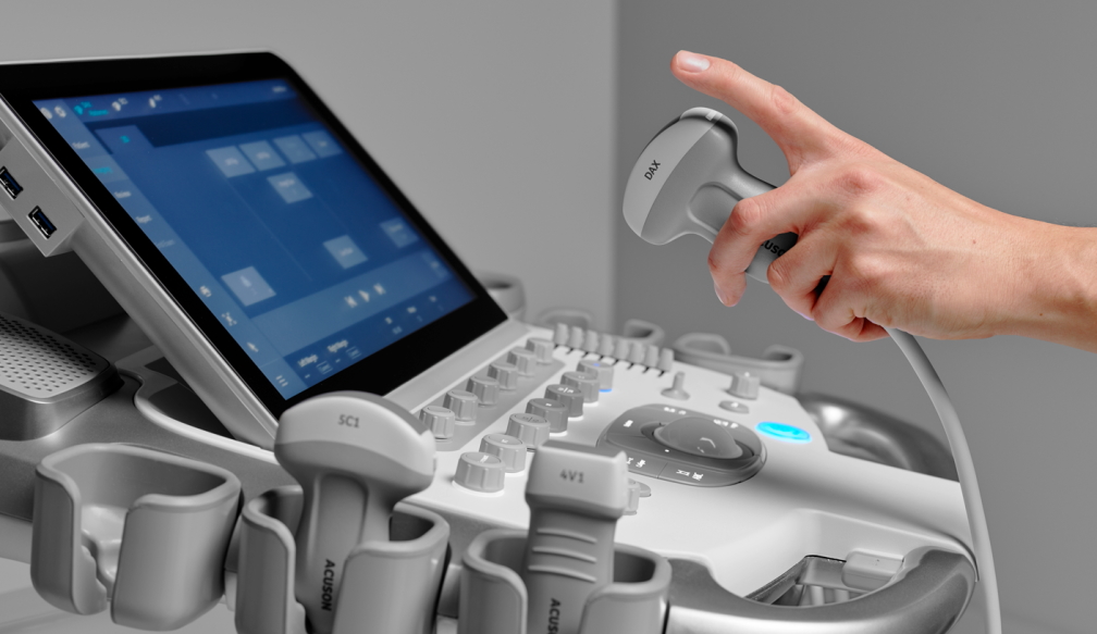
The body's internal structure is visualized by ultrasound imaging, which uses a transducer or probe to generate sound waves. It has no known side effects and produces a clear image of soft tissues that are difficult to see on x-rays. Unexplained pain, edema, and infection are frequently diagnosed with ultrasound. It can also be used to see and assess diseases involving blood flow or to guide needle biopsies with imaging. It is also the most used imaging method for monitoring pregnant women and unborn children. Read more to know how ultrasound is used in healthcare and how it works:
Why is ultrasound used in healthcare?
This is crucial for the examination of fetal and gonadal tissue as ultrasound uses non-ionizing sound waves. Ultrasound has not been linked to carcinogenesis. In most centers, ultrasonography is more widely accessible than more sophisticated cross-sectional modalities like CT or MRI. Ultrasound is easier and less expensive to perform than CT or MRI and has fewer contraindications than those imaging modalities. Both physiology and anatomy can be evaluated with ultrasonography due to their real-time nature. Doppler analysis of the organs and arteries offers a dimension of physiological information that is not present in other modalities. As opposed to MRI or CT, ultrasound is not adversely affected by metallic objects. One can readily extend an ultrasound Narellan examination to examine another organ system or the contralateral extremity.
How do you prepare?
Wear loose-fitting and comfortable clothing for treating ultrasound narellan. All of your clothing and jewelry may need to be taken off in the area to be examined. The procedure would need you to change into a gown. The kind of examination you will have will determine how you should prepare for the procedure. Your doctor can advise you to do fasting for up to 12 hours before some scans. For newborns and young children, this window is smaller. For others, the doctor can advise them to refrain from urinating and drink up to six glasses of water two hours before the exam. By doing this, you can make sure your bladder is full before the scan starts.
How does the procedure work?
The concepts behind sonar used by ships, bats, and fishermen also apply to ultrasound imaging. A sound wave reflects or echoes back when it hits an item. These echo waves can be measured to find the object's distance from us as well as its size, form, and consistency. This includes the object's composition, such as whether it is solid or liquid. In an ultrasonic examination, a transducer transmits sound waves and also captures echoes or returning waves. Small pulses of inaudible, high-frequency sound waves are sent into the body when the transducer is pushed against the skin. The sensitive receiver in the transducer captures minute variations in the pitch and direction of the sound. These distinctive waves are quickly measured by a computer, which then shows them on a monitor as real-time images.
Bottom Line
Your doctor may call you for an ultrasound to examine your organs. Ultrasound helps to better understand one's body and aids in making precision treatments. When you are going for an ultrasonic treatment, be prepared with the instruction given by the medical professionals.


